- ALL COMPUTER, ELECTRONICS AND MECHANICAL COURSES AVAILABLE…. PROJECT GUIDANCE SINCE 2004. FOR FURTHER DETAILS CALL 9443117328
Projects > COMPUTER > 2019 > NON IEEE > APPLICATION
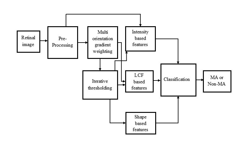
Retinal microaneurysms (MAs) are the earliest clinical sign of diabetic retinopathy disease. Detection of microaneurysms is crucial for the early diagnosis of diabetic retinopathy and prevention of blindness. In this paper, a novel and reliable method for automatic detection of microaneurysms in retinal images is proposed. In the first stage of the proposed method, several preliminary microaneurysm candidates are extracted using a gradient weighting technique and an iterative thresholding approach. In the next stage, in addition to intensity and shape descriptors, a new set of features based on local convergence index filters is extracted for each candidate. Finally, the collective set of features is fed to a hybrid sampling/boosting classifier to discriminate the MAs from non-MAs candidates. The method is evaluated on images with different resolutions and modalities (RGB and SLO) using six publicly available datasets including the Retinopathy Online Challenges dataset (ROC). The proposed method achieves an average sensitivity score on the ROC dataset outperforming state-of-the-art approaches in an extensive comparison. The experimental results on the other five datasets demonstrate the effectiveness and robustness of the proposed microaneurysms detection method regardless of different image resolutions and modalities.
In the existing system, an automatic method to detect microaneurysms in retina photographs. Microaneurysms are the most frequent and usually the first lesions to appear as a consequence of diabetic retinopathy. So, their detection is necessary for both screening the pathology and follow up (progression measurement). Automating this task, which is currently performed manually, would bring more objectivity and reproducibility. To detect them by locally matching a lesion template in sub bands of wavelet transformed images. To improve the method performance, we have searched for the best adapted wavelet within the lifting scheme framework. The optimization process is based on a genetic algorithm followed by Powell’s direction set descent. Results are evaluated on 120 retinal images analyzed by an expert and the optimal wavelet is compared to different conventional mother wavelets. These images are of three different modalities: there are color photographs, green filtered photographs, and angiographs. Depending on the imaging modality, microaneurysms were detected with sensitivity of respectively 89.62%, 90.24%, and 93.74% and a positive predictive value of respectively 89.50%, 89.75%, and 91.67%, which is better than previously published methods.
In the proposed system, A novel method is proposed for accurate and reliable detection of microaneurysms with the possibility of applying this method in large screening setups. The method outperforms state-of-the-art techniques; A new MA candidate extraction technique is employed which extracts the potential MA candidates with a notable improvement in sensitivity; A new set of features based on local convergence index filters is introduced which are then incorporated into a hybrid sampling/boosting classifier. Published two new publicly available retinal image databases, RC-RGB-MA (RGB fundus camera) and RCSLO- MA (scanning laser ophthalmoscope) to evaluate microaneurysms detection methods on different image modalities. Moreover, we provided a software tool for microaneurysm annotation (RC-MAT) helping the experts to collect more labeled Mas
ARCHITECTURE DIAGRAM